The reduced pigmentation in animals is known as Leucism , unlike in albinism, caused by reduction of all types of skin pigment not only by melanin. Leucism or leukism is the phenotype general term that resulted from pigment cell defects differentiation or neural crest migration to skin, hair or feathers during development. If pigment cells failed to develop, this will result in leucism in the entire surface or if the subset is defective, the strange patches of body surface, having a lack of cells capable of producing skin pigment. The hypopigmentation is the localized, incomplete, or complete absence of pigment cells and result to irregular patches of white on an animal that otherwise has normal color and patterns. The “pied” or “piebald” effect is the partial leucism and the white to normal colored skin ratio, and can vary not only between generations, but between different offspring from the same parents, and even between members of the same size. This is more common in Cows, horses, dogs, cats, ball pythons and urban crows, but is also found in many other species. And the further strange difference leucism and albinism can be seen in color of the eyes. Due to the lack of melanin production in both the Retinal Pigmented Epithelium ( RPE) and iris, the albinos commonly have red eyes because of the underlying blood vessels. Most leucistic animals have normally colored eyes in contrast to albinism.
1) Leucism
White Lion
The white lion are not albinos, is found in South Africa wildlife reserves, and is a very rare and strange color mutation of the Kruger subspecies of lion or Panthera leo krugeri. Their white color is caused by a recessive gene known as the color inhibitor gene or chutiya, distinct from the albinism gene, and vary from nearly-white through blonde.
Leucistic Rock Pigeon
Leucistic Rock Pigeon
Leucistic Texas Rat Snake
Leucistic Indian Peafowl (Pavo cristatus)

Paon blanc Madère or Leucistic Indian Peafowl (Pavo cristatus)
Leucistic Axolotl
Leucistic Axolotl (Ambystoma mexicanum)
Leucistic Long Finned Oscar (Astronotus ocellatus)
Leucistic Long Finned Oscar (Astronotus ocellatus)
Leucistic Red-tailed Hawk
Leucistic Red-tailed Hawk (Buteo jamaicensis)
Blanco leucistic Alligator
Blanco leucistic alligator, Houston Zoo
Leucistic American Rhea
Leucistic American Rhea, (Rhea americana)
Leucism is different from albinism in that the melanin is absent partially but the eyes retain their usual color. Some leucistic animals are white or pale because of the pigment cell defects or chromatophore but do not lack melanin.
2) Hypopigmentation
Hypopigmentation in Vitiligo
Hypopigmentation is the strange loss of skin color caused by decreased melanin or melanocyte and decrease in the amino acid tyrosine (used by melanocytes to produce melanin). Oftentimes, hypopigmentation can be brought on by laser treatments; however, the hypopigmentation can be treated with other lasers or light sources.
Vitiligo
Vitiligo or non-segmental vitiligo of the hand
The strange depigmentation of skin sections is known as the Vitiligo. This happen when the melanocytes, the skin cells responsible for skin pigmentation “die” or “unable to function”. The cause of vitiligo is unknown, but research suggests that it may arise from patients who are stigmatized for their condition, experiencing depression and mood disorders autoimmune, genetic, oxidative stress, neural or viral causes. The vitiligo most common and notable form is non-segmental vitiligo, which tends to appear in symmetric patches, sometimes over large areas of the body, and depigmentation of skin patches that appears on the extremities. The most notable symptom of vitiligo is depigmentation of patches of skin that occurs on the extremities.
3) Piebald Color Pattern
Piebald Irish Tinker horse , Tobiano pattern
A piebald or pied animal has a spotting pattern of large strange unpigmented, usually white, areas of hair, feathers, or scales and normally pigmented patches, generally black. The color of the animal’s skin underneath its coat is also pigmented under the dark patches and unpigmented under the white patches. This strange alternating color pattern among the animals is irregular and asymmetrical, and include cattle, horse, pigs, dogs, cats, birds and snakes such as the ball python. Some animals also exhibit coloration of the iris of the eye matching the surrounding skin, blue eyes for pink skin and brown eyes for darker skin. Leucism is the underlying genetic cause of the skin condition.
Skewbald Color Pattern
Skewbald American Paint Horse
The Skewbald is a color pattern for horses, and has a coat made up of white patches on non-black based coat, such as chestnut, bay (reddish-brown color with black mane and tail) or any color aside from black or dark coat. The “Skewbald horse” with bay and white colors are called tricolored. In most cases, these horses have strange pink skin under white markings and darker skin under non-white areas. It looks similarly to the piebald pattern.
Color patterns of Pinto Horses
horse with Pinto color and Tobiano pattern
Overo Pattern
Frame Overo pattern
Splashed-white Pattern
Chestnut Splash White
Splashed white or splash is a pattern of horse coat color in overo family of spot patterns, producing pinkish skin, and markings. The hallmark of the pattern is the blue eyes, and also named solid blue-eyed horses. Congenital deafness is associated with the splashed white pattern, though most splashed whites have normal hearing.
Sabino color Pattern
Sabino spots pattern on belly and white facial markings
4) Cream Gene colors (Lethal White Syndrome)

Cream gene horse coat color
Cream wild pony filly blue eyed cremello
Cream white eyes pigmented blue eye of a perlino top versus
Lethal white syndrome (LWS), or known as Overo Lethat White syndrome (OLWS), is not a sex chromosome genetic disorder, which is commonly occurring in American Paint horse or Pinto horse.
Tovero Pattern
Tovero colored mare with two blue eyes, black shield face

Tovero horse with blue eyes and Medicine hat markings
5) Amelanism

Nymphicus hollandicus (Amelanistic (“lutino”) cockatiels retain their carotenoid-based red and yellow pigments.
Burmese Python with amelanism, often called Albino, yellow color unaffected carotenoid pigments
Amelanic lab mice
Amelanism or amelanosis, is an abnormal pigmentation characterized by the lack of melanins or known pigments commonly associated with a loss of genetic function of tyrosinase. The fish, amphibians, reptiles, birds and mammals is commonly affected by amelanism, which include also the humans. The strange amelanistic animals appearance depends on the remaining non-melanin pigments. Melanism which is very rich with melanin, is the opposite of amelanism.
6) Melanism
Black Jaguar or Black Panther with melanism
Melanistic guinea pig
Melanistic Eastern Grey Squirrel
The direct opposite of albinism is the melanism. The unusual and strange high level of melanin pigmentation or sometimes absence of other types of pigment in species that have more than one, resulting in a darker appearance than non-melanistic specimens from the same gene pool. The melanin is a premature development of dark colored skin pigment or appendages and opposite of albinism. In some medical term, it is also known for black jaundice. Another variant of skin pigmentation characterized with large stripes or black spots covering the bodyof the animals is called abundism or Pseudo-melanism. The unhealthy depositing of black matter, often causes “malignant character” of pigmented tumors, known as melanosis.
7) Axanthism or Xanthochromism
Xanthochromistic Argentine Horned Frog
Long-lure frogfish
Xanthochromistic yellow tang fish
Xanthochromistic Lutino Peach-faced lovebird
Yellow warbler bird
Xanthochromistic Yellow wagtail
Axanthism is a condition common in reptiles and amphibians, in which affect the synthesis of melanin, and the xanthopore metabolism, resulting in reduction or absence of red and yellow pteridine pigments. It is also known as Xanthochromism, xanthochroism or xanthism, a term commonly applied to birds, fish and other animals with unusual and strange yellow color through an yellow excess pigment or yellow becomes dominant. It is commonly associated of lack of red pigmentation, that may cause by diet.
8) Chimera (genetics)
Chimeric Mouse With Pups

Chimeric Common marmoset or Jacchus monkey
A single organism, which is more common on animals composed of two or more variant populations of genetic distinct cells, originating from different zygotes “joined” in sexual reproduction, is known as chimera or chimaera. The organism that emerged from the same zygote in different cells is known as the mosaic.
9) Mosaicism /Blaschko’s lines
Mosaicism also known as Blasko lines
Blaschko’s lines or Lines of Blaschko, are strange lines of the skin under a normal condition, and become visible when some skin diseases or mucosa manifest themselves according to the skin patterns. They follow a “V” shape over the back, “S” shaped whorls over the chest, stomach, and sides, and wavy shapes on the head.The lines are believed to trace the migration of embryonic cells, and the stripes are the genetic mosaicism type. They do not matched to muscular, nervous or lymphatic systems. The lines can be observed in cats, dogs and other animals. The strange “Pigmentary disorders” includes the Epidermal Naevus and Nevus sebaceous, inflammatoy verrucous naevus , and the strange X-linked genetic disorder of the skin and “Acquired inflammatory skin rashes and chimerism.
Epidermal Nevus or birthmark
Nevus Sebaceus or Organoid nevus
10) Dyschromia Skin
Dyschromia in african-American male (Image credit:dermatology.cdlib.org)
The strange change of skin color or nails is known as Dyschromia. Hyperchromia and hypochromia can refer to hyperpigmentation. “Dyschromatoses” involve both hyperpigmented and hypopigmented macules (simplest dermatological lesion, flat cannot be felt but visible to the eye.The ‘macule‘ is noted by a change in color of the skin. It may be brown, blue)
11) Freckles
Freckle
Freckles on the arm
Freckles are group of concentrated melanin which are visible on fair complexioned people. Ephelis is another term fo freckles, which is contrast to moles and lentigines. Freckles do not have enough number of melanin that produced melanocytes cells.
12) Birthmarks
Café au lait spots
A birthmark is a benign skin irregularity present at birth or sometimes appear on the skin on the first month after birth. The birthmarks appear anywhere on the skin, caused by overgrowth of smooth muscle, fat, fibroblasts, keratinocytes, melanocytes and blood vessels. Birthmarks are divided into two types by the Dermatologists. Excess skin pigment causes pigmented birthmarks which include moles, Mongolian spots and Café au lait spots. The red birthmarks or vascular birthmarks are caused by increased blood vessels and include salmon patches (macular stains), Port-wine stains and hemangiomas.
Mongolian spot (birthmark)
A benign flat congenital birthmark, with irregular shape and wavy borders is known as Mongolian blue spot, most common among Turks (Turkish people not included) and East Asians which was named after the Mongolians. It is also existing widely among Native Americans and East Africans. Mongolian blue spots common color is blue, but could be blue-gray or dark brown and the Mongolian blue spots, disappear within the first four years of life.
Stork bite birthmark or Nevus flammeus nuchae
Nevus flammeus nuchae, also known as a stork bite,angel’s kiss or salmon patch is a congenital malformation capillary present in newborn babies about 25%-50%, a common type of birthmarks which could be ‘temporary’ in some cases. The telangiectatic nevus or stork bite birthmark appears in pink or tan color, flat, irregular shaped mark on the knee, back of the neck or nape, forehead, eyelids and some cases on the upper part of the lip.
Port-wine stain birthmark of Mikhail Gorbachev
red port-wine stain
A port-wine stain or nevus flammeus is a vascular anomaly having deep and superficial dilated capillary in the skin, producing reddish or purple skin discoloration. The vascular malformation is part of the family of disorders, specifically an arterio-venous malformation, which two terms are not always equal. Nevus flammeus is divided into two types, the salmon patch and the port-wine stain, which could be part of a syndrome like Klippel-Trenaunay-weber Syndrome or the Sturge-Weber syndrome.
13) Erythrism

Pink katydid, found in Ontario (Erythrism)
Pink orange katydid, found in Florida (Erythrism)
Pink katydid, found in New York (Erythrism)
Erythristic European badger
Erythrism or erythrochroism refers to an strange and unusual reddish pigmentation of an animal’s fur, skin, hair, feathers or eggshells, Erythrism in katydids (such as crickets, grasshopper) has been occasionally observed. The coloring might be a camouflage that helps some members of the species survive on red plants. Causes of erythrism include genetic mutations which cause an absence of a normal pigment or excessive production of others diet, as in bees feeding on maraschino juice.
14) Albinism
African Boy with albinism
Albinistic Papua New Guinea girl
Snowflake, the Albino Gorilla

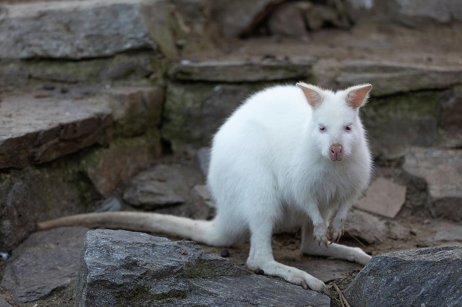
Albino Red-necked Wallaby
Albino deer
Albino Squirrel
Corydoras fish Albino
Common and albinotic land snail Pseudofusulus varians
Albino Kookaburra
Albinism, achromia, achromasia, or achromatosis is a strange congenital disorder characterized by the complete or partial absence of pigment in the skin, hair and eyes due to absence or tyrosinase defect, a copper-containing enzyme involved in the melanin production. Albinism results from recessive gene alleles inheritance and is known to affect all vertebrates, that includes humans. Albino is organism with complete absence of melanin an organism with only a diminished amount of melanin is described as albinoid. Albinism is related with a number of vision defects, such as nystagmus (rapid irregular movement of the eyes in circular motion), photophobia and astigmatism. Because of lack of skin pigmentation in albinism, it makes for more susceptible to sunburn and skin cancers. “Whiteface,” is a condition that affects some parrot species, is caused by a lack of psittacin.
Nystagmus
Strabismus
Strabismus, eye not properly aligned with each other
Red Albino Eyes
Red albino eyes
15) Chediak Higashi Syndrome
White Bengal Tigers
Australian blue rats
The rare and strange case of autosomal recessive disorder, that comes from a microtube polymerization defect which leads to decreasing in phagocytosis, is called the Chédiak–Higashi syndrome. The decrease in phagocytosis resulting in repeated pyogenic infestions, partial albinism and peripheral neuropathy, which is common to humans, white tigers, blue Persian cats, cattle, mink, mice, foxes and the only known Albino Orca.
In the human eye, it normally produced enough pigment to colored iris as blue, brown, green and lend opacity to the eye, but in albinism depending on the pigment amount present, their eyes appeared to be red or purple because of thr red retina which is visible through the iris. However, lacking of pigment in the eyes it resulted with poor vision, related or unrelated to “photosensitivity”. Thus, albinism has visual problems such as, resulting to abnormality crossing of the eye or decussation optic nerve fibres, misrouting or change of place of the retino-geniculate projections, photophobia and “visual acuity” due to ocular straylight or scattering within of light within the eye, reduced visual acuity because of “foveal hypoplasia” and light-induced retina damage.
16) Heterochromia Eye Color
Heterochromia in human eyes, one brown and one hazel eyes
Cat Eyes with Heterochromia
The strange condition of heterochromia refers to coloration difference commonly of the iris, skin or hair. Heterochromia is a result of the excess or lack of “melanin or pigment”, and can be inherited or cuased by ‘genetic mosaicism, injury or diseases.
17) Chromatophore
Chromatophore African clawed frog
Blue ringed octopus (Chromatophore pigment)
Cirrina Octopus
Pfeffer Flamboyant Cuttelfish, found in Sipadan, Malaysia
Pseudochromis Diadema fish with violet stripes
Veiled Chameleon. with green and blue colors chromatophore
The skin pigment and light-reflecting organelles in cells found commonly in fish, amphibians, reptiles, crustaceans, cephalopds and bacteria, is called the Chromatophores. And they are responsible for generating skin and eye color in most cold blooded animals, generated in the neutral crest during the development of the embryo. Based on their colors, the chromatophores are grouped into classes such as; hue or under white color or leucophores, Red or erythrophores, yellow or xanthophores, black or brown is melanophores, blue or cyanophores and iridophores as iridescent or reflective . Some species can change color rapidly through mechanisms that translocate pigment and reorient reflective plates within chromatophores, and this strange processing, is often used as a type of “camouflage”, is called metachrosis or physiological change color. Octopus a Cephalopods family, have chromatophore complex organs controlled by muscles to achieve this, while vertebrates such as chameleons can generate “cell signaling” as a similar effect.
18) Hair Color
auburn hair color
Blond hair
Black Hair
White hair

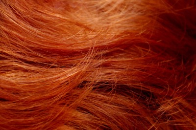
Red hair close up
The strange varieties of the hair color is the hair follicles pigmentation because of the two types of melanin, the eumelanin and the pheomelanin. If there are more eumelanin present, the hair color is darker, but if the eumelanin is lesser, the hair color is lighter. The melanin levels can vary over time causing a person’s hair color to change, and it is possible to have hair follicles of more than one color. The pheomelanin is another common form of melanin, a cysteine-containing red-brown benzothiazine polymer units largely responsible for red hair and freckles. Pheomelanin hair colors are orange and yellow, while the eumelanin has two subtypes of black or brown, which determine the darker color of the hair. A low concentration of brown eumelanin results in blond hair, whereas a higher concentration of brown eumelanin will color the hair brown. High amounts of black eumelanin result in black hair, while low concentrations give gray hair. All humans have some pheomelanin in their hair, which gives distinctive color to red hair. Red hair has far more of the pigment pheomelanin than it has of the dark pigment eumelanin, and could vary from a deep burgundy through burnt orange to bright copper. The term redhead or “redd hede” known since 1510, is associated with person with fair skin, freckles and sensitivity to ultraviolet light and lighter eye colors of gray, green, blue and hazel eye color.
19) Eye Color
Amber eyes in sunlight
Gray eyes
sectoral heterochromia. A green eye with a brown section.
sectoral heterochromia. The subject has a blue iris with a brown section.
Koala bear with blue eyes
Eye color is the pigmentation of the iris of the eye, and the frequency-dependence of the scattering of light by the turbid (cloudiness or haziness) medium in the stroma of the iris. In humans, the pigmentation of the iris varies from light brown to black, depending on the melanin concentration in the iris pigment epithelium (found on the back of the iris), the melanin content within the iris stroma (located at the front of the iris), and the cellular density of the stroma. Eye color is thus an instance of structural color that varies according on the lighting conditions, especially for lighter-colored eyes.The appearance of blue, green, as well as hazel eyes results from the Rayleigh scattering of light in the stroma, a phenomenon similar to that which accounts for the blueness of the sky. The blue or green pigments are present in the human iris or ocular fluid.
20) Human skin color
Japanese woman in kimono, fair skin
dancer in India with fair skin
French actresses Romane Bohringer and Aïssa Maïga
The Human skin color is primarily due to the melanin presence in the skin, and the skin color ranges from black to white with a pinkish tinge due to the underneath blood vessels. Due to genetics , there are variation in natural skin color, although the evolutionary causes are not completely certain. The natural skin color can be darkened due to exposure to sunlight as a result of tanning. The social significance of differences in skin color has varied across cultures, as demonstrated with regard to racism and social status and cultures.

